Endobronchial Ultrasound (EBUS)
Endobronchial ultrasound (EBUS) is a minimally invasive but highly effective procedure used to diagnose lung cancer, infections, and other diseases causing enlarged lymph nodes in the chest. UC San Diego Health was one of the first centers on the West Coast to offer EBUS and remains a regional leader in volume and overall experience.
Why is EBUS used?
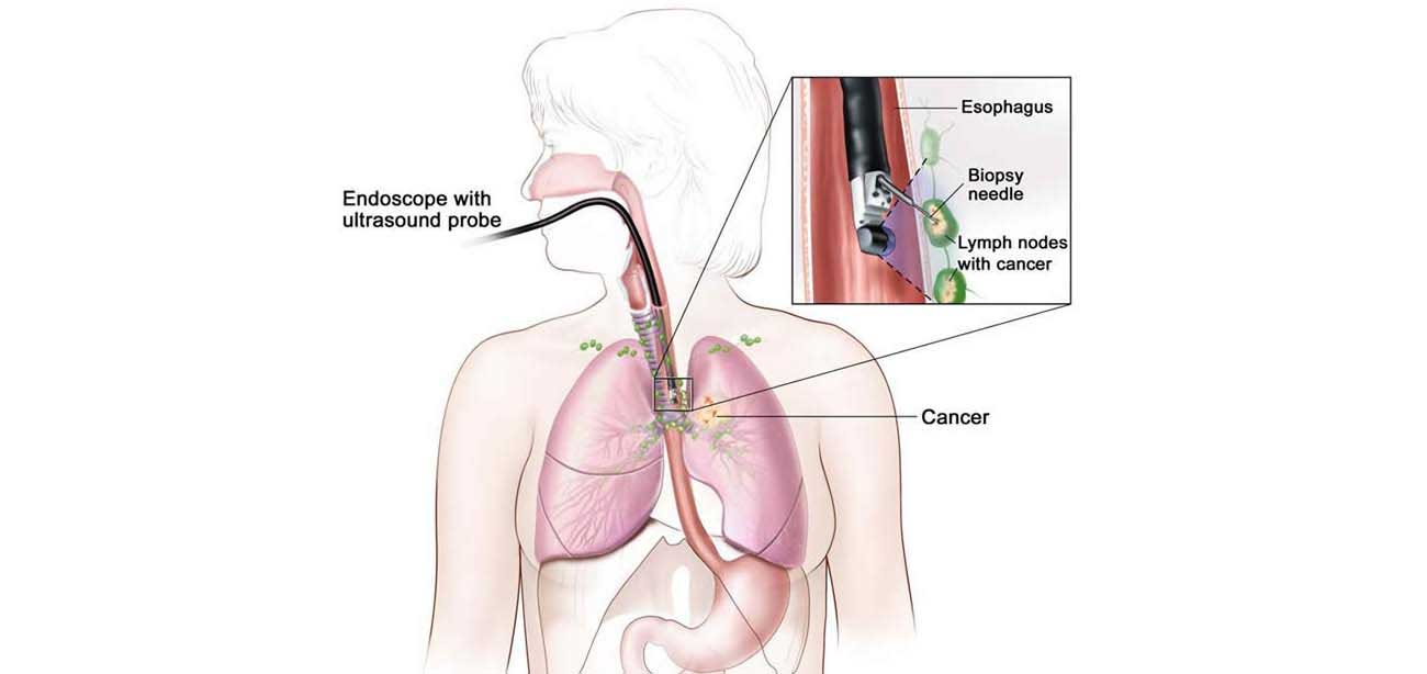
EBUS allows physicians to perform a technique known as transbronchial needle aspiration (TBNA) to obtain tissue or fluid samples from the lungs and surrounding lymph nodes without conventional surgery. The samples can be used for diagnosing and staging lung cancer, detecting infections, and identifying inflammatory diseases that affect the lungs, such as sarcoidosis or cancers like lymphoma.
What makes EBUS different?
During a conventional diagnostic procedure, surgery known as mediastinoscopy is performed to provide access to the chest. A small incision is made in the neck just above the breastbone or next to the breastbone. Next, a thin scope, called a mediastinoscope, is inserted through the opening to provide access to the lungs and surrounding lymph nodes. Tissue or fluid is then collected via biopsy.
An endobronchial ultrasound is much less invasive.
 Call For Appointment